Thursday 15 May 2014

Basics Of ELECTRENCEPHALOGRAM (EEG)

An electroencephalogram is a recording of brain activity.
Brain cells continually send messages to each other that can be picked up as small electrical impulses on the scalp. The process of picking up and recording the impulses is known as an EEG.
A normal EEG means that you have a normal pattern of brainwave activity. An abnormal reading means that abnormal patterns of brain activity are being produced and picked up.
People of all ages can have EEGs, including tiny babies and the very elderly.

Epilepsy

If you have epilepsy, your brain sometimes doesn't work normally. This causes seizures (previously known as epileptic fits).
People with seizures can have normal brain activity (EEG results) or quite minor changes in between attacks. This is why specialists are needed to look at and assess your EEG results.
An EEG will help your doctor identify the type of epilepsy you have and what may be triggering your seizures. This will enable the most effective type of medication to be prescribed for you.
In rare cases, treatment may include neurosurgery (brain surgery).

Other conditions

Other conditions that affect brain function where EEGs can be used include:
  • dementia - a group of symptoms that are responsible for the decline of brain function
  • head injury
  • brain tumour - an abnormal and uncontrollable growth of cells in the brain
  • brain abscess - a pus-filled swelling in the brain that's caused by an infection
  • encephalitis - brain inflammation that's sometimes caused by an infection
  • brain haemorrhage - bleeding in the brain
  • cerebral infarct - brain tissue that has died due to a blockage in blood flow
  • sleep disorders
  • coma

How EEG is performed

Routine EEG recordings usually take 20-40 minutes (see below), although a typical appointment will last for about an hour including some preparation time at the beginning plus some time at the end.
The test is usually carried out as an outpatient procedure by a highly trained clinical physiologist who has specialised in neurophysiology (the study of the workings of the nervous system).
The clinical physiologist will explain the procedure to you, and you’ll be able to ask any questions that you have. You'll also be asked to agree or decline to have the various parts of the test (consent). The EEG procedure is painless and you should feel comfortable throughout.
The skin on your scalp will be cleansed and about 20 electrodes (small discs) will be attached to specific areas, measured out in the correct locations.
The electrodes will be connected to an EEG machine by thin leads. The machine records your brain wave activity for later analysis.  Afterwards, the electrodes will be removed and your scalp cleaned. You may also want to wash your hair afterwards
After you’ve left the EEG department, the recordings will be analysed and used for the purposes of diagnosis and  treatment.

Types of EEG

The main types of EEG are explained below.

Routine EEG

A routine EEG recording lasts for about 20-40 minutes.
During the test, you’ll be asked to rest quietly and from time to time to open or close your eyes. In most recordings you’ll be asked to breathe deeply in and out for about three minutes.
At the end of the procedure, provided you've agreed, a strobe light will be placed nearby and you'll see bright flashes of light which are repeated at different speeds.
The reasons for doing this and your consent will always be clearly established beforehand.

Sleep EEG

A sleep EEG is carried out while you're asleep. It may be used if a routine EEG doesn't show any conclusive features, or to test for sleep disorders.
While you’re asleep your brainwave patterns change significantly, and useful information related to your condition can be obtained. If necessary, to promote sleep, you may be asked to stay awake during the preceding night.

Ambulatory EEG

An ambulatory EEG is where brain activity is recorded throughout the day and night, over a period of one or more days.
You’ll be given a small portable EEG recorder that can be clipped onto your clothing. It will record your EEG activity during the whole day and night.

Video telemetry

Video telemetry, also known as video EEG, is a special type of EEG that simultaneously videos you and records your brain wave activity.
It’s used when an EEG and continuous intensive monitoring are needed. For example, it can be used to see what a child is doing while they’re having a seizure. This can help diagnose the type of epilepsy that they have, where the seizure starts and how the electrical activity spreads through their brain.
Video telemetry is usually carried out on an in-patient basis in a purpose built hospital suite. It usually takes place day and night for up to five days, unless enough information about the seizure is recorded over a shorter period.
The EEG signals are transmitted wirelessly to a base-station (a computer processing unit). The video is both recorded at the base-station and kept under regular surveillance by trained staff so they can respond immediately if there are any problems.

Tuesday 6 May 2014

BRAIN DISORDERS..SIMPLE ANALYSIS

    Overview

    Your brain is the control center of your body. It is a part of the neurological system, a complex system that includes the spinal cord and a vast network of nerves and neurons that control and implement the functions you do every day. Brain disorders occur when your brain is damaged by injury, disease, or health conditions.

    What Are the Symptoms of Brain Disorders?

    The symptoms of brain disorders typically depend on the cause of the condition. Brain disorders may affect the main areas of your brain that control how you move, think, and behave. Some tumors can constrict the blood vessels in your brain.
    The following are some common symptoms brain disorders may present:
    • confusion or problems concentrating
    • headaches or migraines
    • seizures (convulsions)
    • memory problems
    • change in the way you normally behave
    • problems with your vision (double vision, for example)
    • lack of muscle control
    • vomiting or nausea

    What Causes Brain Disorders?

    The causes of brain disorders vary with the type of disorder you experience
    The following are causes of brain disorders:
    • trauma to the brain
    • stroke (restricted or reduced oxygen and blood in the brain that leads to cellular death)
    • viral infections (viruses may cause inflammation and swelling in the brain’s tissue)
    • disease and cancer
    • abnormal growths (tumors)
    • inherited conditions that affect the brain
    • change in your brain’s electrical pathways (communication between neurons)

    Who Is at Risk For Brain Disorders?

    You may be at risk for a brain disorder if you:
    • have blunt trauma to the head
    • have a family history of brain disorders or disease
    • have a viral infection
    • have a stroke
    • smoke tobacco products
    • stop breathing (can prevent oxygen from reaching the brain)

    Types of Brain Disorders

    There are many types of brain disorders, and they can change the way your brain commands the rest of your body.

    Brain Injuries

    Brain injuries are often caused by blunt trauma. Injury can damage tissue, neurons (messengers within the brain), and nerves that transmit information from the brain to your body. This can cause changes in how your brain communicates with the rest of your body.

    Brain Tumors

    Tumors can develop in the brain’s tissue and cause many problems, including preventing blood circulation in the brain. These growths may be cancerous or benign.

    Degenerative Diseases

    Degenerative diseases can affect the brain in many ways. They can change your personality, cause confusion, or destroy your brain’s tissue and nerves. Some brain diseases, such as Alzheimer’s disease, may manifest as you age and slowly impair your memory and thought processes. Other diseases, such as Tay-Sachs disease, begin at an early age. Tay-Sachs disease affects a child’s mental and physical capabilities.

    Mental Health Conditions

    Mental health conditions change your behavior patterns. Certain types of mental health conditions may be chronic or acute. Depressionanxiety, and bipolar disorder are three brain disorders that may become chronic conditions.

    Diagnosing Brain Disorders

    Your primary physician may refer you to a specialist in the neurological field. This specialist may perform a neurological exam to check your vision, hearing, and balance.
    The doctor might also use imaging technology—such as a computed tomography (CT) scan—to take images of your brain. Other diagnostic imaging tools include magnetic resonance imaging (MRI) and positron emission tomography (PET).
    In addition, your neurologist might take and study fluid from your brain and spinal cord as a way to locate bleeding in the brain, infection, and other abnormal occurrences.

    Treating Brain Disorders

    Treatment is based on the doctor’s findings, diagnosis, and your overall health. Your doctor might combine treatments to improve your condition.

    Medication

    If you have swelling or inflammation in your brain, medications to reduce these symptoms may be used.
    For mental health and mood disorders, such as depression, psychotropic drugs may be prescribed to control your behavior.
    For degenerative conditions that cause the loss of muscle control and movement, drugs that help decrease the symptoms may be options.

    Surgery

    Surgery may be used to remove a brain tumor or damaged tissue or to drain excess fluid caused by infection. Sometimes brain surgery is done to remove a sample of brain tissue or a tumor for diagnostic purposes. The samples are examined for cancer, disease, and other abnormal findings.

Wednesday 30 April 2014

Brachial Plexus brief anatomy

The brachial plexus is a network of nerve fibers, running from the spine, formed by the ventral rami of the lower four cervical and first thoracic nerve roots (C5-C8, T1). It proceeds through the neck, the axilla (armpit region), and into the arm. It is a network of nerves passing through the cervico-axillary canal to reach axilla and innervates brachium (upper arm), antebrachium (forearm) and hand

Function

The brachial plexus is responsible for cutaneous and muscular innervation of the entire upper limb, with two exceptions: the trapezius muscle innervated by the spinal accessory nerve (CN XI) and an area of skin near the axilla innervated by the intercostobrachial nerve.
Lesions can lead to severe functional impairment.[1]

Anatomy

The brachial plexus is divided into Roots, Trunks, Divisions, Cords, and Branches. There are five "terminal" branches and numerous other "pre-terminal" or "collateral" branches that leave the plexus at various points along its length.

Root

The five roots are the five anterior rami of the spinal nerves, after they have given off their segmental supply to the muscles of the neck. The brachial plexus emerges at five different levels; C5, C6, C7, C8, and T1. There is prefixed or postfixed formations in some cases which involves c4 or T2 respectively .

Trunk

These roots merge to form three trunks:

Division

Each trunk then splits in two, to form six divisions:
  • anterior divisions of the upper, middle, and lower trunks
  • posterior divisions of the upper, middle, and lower trunks

Cord

These six divisions will regroup to become the three cords. The cords are named by their position with respect to the axillary artery.
  • The posterior cord is formed from the three posterior divisions of the trunks (C5-C8,T1)
  • The lateral cord is the anterior divisions from the upper and middle trunks (C5-C7)
  • The medial cord is simply a continuation of the anterior division of the lower trunk (C8,T1)

Branches

The branches are listed below. Most branch from the cords, but a few branch (indicated in italics) directly from earlier structures. The five on the left are considered "terminal branches".

Diagram

Dorsal scapular nerve (rhomboids, levator scapulae) Suprascapular nerve (supraspinatus, infraspinatus) Nerve to subclavius (subclavius) Lateral pectoral nerve (pectoralis major) Musculocutaneous nerve (coracobrachialis, brachialis, biceps brachii) Axillary nerve (deltoid, teres minor) Median nerve (forearm flexors except FCU and ulnar part of FDP, thenar muscles) Ulnar nerve (FCU and ulnar part of FDP, most intrinsic hand muscles Medial cutaneous nerve of forearm Medial cutaneous nerve of arm Radial nerve (triceps brachii, supinator, anconeus, forearm extensors, brachioradialis) Lower subscapular nerve (lower part of subscapularis, teres major) Thoracodorsal nerve (latissimus dorsi) Medial pectoral nerve (pectoralis major, pectoralis minor) Upper subscapular nerve (upper part of subscapularis) Long thoracic nerve of Bell (serratus anterior) Cervical spinal nerve 5 Cervical spinal nerve 6 Cervical spinal nerve 7 Cervical spinal nerve 8 Thoracic spinal nerve 1
Anatomical illustration of the brachial plexus with areas of roots, trunks, divisions and cords marked. Clicking on names of branches will link to their Wikipedia entry.
Diagrammatic representation of the brachial plexus using colour to illustrate the contributions of each nerve root to the branches

Specific branches

Bold indicates primary spinal root component of nerve. Italics indicate spinal roots that frequently, but not always, contribute to the nerve.
From Nerve Roots[2] Muscles Cutaneous
roots dorsal scapular nerve C4, C5 rhomboid muscles and levator scapulae -
roots long thoracic nerve C5, C6, C7 serratus anterior -
roots branch to phrenic nerve C5 Diaphragm -
upper trunk nerve to the subclavius C5, C6 subclavius muscle -
upper trunk suprascapular nerve C5, C6 supraspinatus and infraspinatus -
lateral cord lateral pectoral nerve C5, C6, C7 pectoralis major and pectoralis minor (by communicating with the medial pectoral nerve) -
lateral cord musculocutaneous nerve C5, C6, C7 coracobrachialis, brachialis and biceps brachii becomes the lateral cutaneous nerve of the forearm
lateral cord lateral root of the median nerve C5, C6, C7 fibres to the median nerve -
posterior cord upper subscapular nerve C5, C6 subscapularis (upper part) -
posterior cord thoracodorsal nerve (middle subscapular nerve) C6, C7, C8 latissimus dorsi -
posterior cord lower subscapular nerve C5, C6 subscapularis (lower part ) and teres major -
posterior cord axillary nerve C5, C6 anterior branch: deltoid and a small area of overlying skin
posterior branch: teres minor and deltoid muscles
posterior branch becomes upper lateral cutaneous nerve of the arm
posterior cord radial nerve C5, C6, C7, C8, T1 triceps brachii, supinator, anconeus, the extensor muscles of the forearm, and brachioradialis skin of the posterior arm as the posterior cutaneous nerve of the arm
medial cord medial pectoral nerve C8, T1 pectoralis major and pectoralis minor -
medial cord medial root of the median nerve C8, T1 fibres to the median nerve portions of hand not served by ulnar or radial
medial cord medial cutaneous nerve of the arm C8, T1 - front and medial skin of the arm
medial cord medial cutaneous nerve of the forearm C8, T1 - medial skin of the forearm
medial cord ulnar nerve C8, T1 flexor carpi ulnaris, the medial two bellies of flexor digitorum profundus, the intrinsic hand muscles except the thenar muscles and the two most lateral lumbricals the skin of the medial side of the hand and medial one and a half fingers on the palmar side and medial two and a half fingers on the dorsal side
Some mnemonics for remembering the branches:
  • Medial Cord Branches
  • 5 main nerves of brachial plexus, in order laterally to medially
    • "My Aunty Recognised My Uncle" - Musculocutaneous, axillary, radial, median, ulnar.

Injuries

Brachial plexus injury affects cutaneous sensations and movements in the upper limb. They can be caused by stretching, diseases, and wounds to the lateral cervical region (posterior triangle) of the neck or the axilla. Depending on the location of the injury, the signs and symptoms can range from complete paralysis to anesthesia. Testing the patient's ability to perform movements and comparing it to their normal side is a method to assess the degree of paralysis. A common brachial plexus injury is from a hard landing where the shoulder widely separates from the neck (such as in the case of motorcycle accidents or falling from a tree). These stretches can cause ruptures to the superior portions of the brachial plexus or avulse the roots from the spinal cord. Upper brachial plexus injuries are frequent in newborns when excessive stretching of the neck occurs during delivery. Studies have shown a relationship between birth weight and brachial plexus injuries; however, the number of cesarean deliveries necessary to prevent a single injury is high at most birth weights.[3] For the upper brachial plexus injuries, paralysis occurs in those muscles supplied by C5 and C6 like the deltoid, biceps, brachialis, and brachioradialis. A loss of sensation in the lateral aspect of the upper limb is also common with such injuries. An inferior brachial plexus injury is far less common, but can occur when a person grasps something to break a fall or a baby's upper limb is pulled excessively during delivery. In this case, the short muscles of the hand would be affected and cause the inability to form a full fist position.[4] In order to differentiate between a pre ganglionic and a post ganglionic type of injury on clinical examination one has to keep the following points in mind. In pre ganglionic injuries there will be loss of sensation above the level of clavicle, presence of pain in an otherwise insensate hand, presence of ipsilateral Horner's syndrome and loss of function of muscles supplied by branches arising directly from roots i.e. long thoracic nerve palsy leading to winging of scapula and elevation of ipsilateral diaphragm due to phrenic nerve palsy.
Acute brachial plexus neuritis is a neurological disorder that is characterized by the onset of severe pain in the shoulder region. Additionally, the compression of cords can cause pain radiating down the arm, numbness, paresthesia, erythema, and weakness of the hands. This kind of injury is common for people who have prolonged hyperabduction of the arm when they are performing tasks above their head.

Tuesday 29 April 2014

BRAINSTEM OVERVIEW

In the anatomy of humans and of many other vertebrates, the brainstem (or brain stem) is the posterior part of the brain, adjoining and structurally continuous with the spinal cord. In humans it is usually described as including the medulla oblongata (myelencephalon), pons (part of metencephalon), and midbrain (mesencephalon).[1][2] Less frequently, parts of the diencephalon are included. The brainstem provides the main motor and sensory innervation to the face and neck via the cranial nerves. Of the twelve pairs of cranial nerves, ten pairs come from the brainstem. Though small, this is an extremely important part of the brain as the nerve connections of the motor and sensory systems from the main part of the brain to the rest of the body pass through the brainstem. This includes the corticospinal tract (motor), the posterior column-medial lemniscus pathway (fine touch, vibration sensation, and proprioception), and the spinothalamic tract (pain, temperature, itch, and crude touch). The brainstem also plays an important role in the regulation of cardiac and respiratory function. It also regulates the central nervous system, and is pivotal in maintaining consciousness and regulating the sleep cycle. The brainstem has many basic functions including heart rate, breathing, sleeping, and eating.

Sunday 13 April 2014

Neurotransmittes- A Review and its types..

Neurotransmitters are endogenous chemicals that transmit signals across a synapse from one neuron (brain cell) to another 'target' neuron.[1] Neurotransmitters are packaged into synaptic vesicles clustered beneath the membrane in the axon terminal, on the presynaptic side of a synapse. Neurotransmitters are released into and diffuse across the synaptic cleft, where they bind to specific receptors in the membrane on the postsynaptic side of the synapse.[2] Many neurotransmitters are synthesized from plentiful and simple precursors, such as amino acids, which are readily available from the diet and which require only a small number of biosynthetic steps to convert.
Most neurotransmitters are about the size of a single amino acid, but some neurotransmitters may be the size of larger proteins or peptides. A neurotransmitter is available only briefly – before rapid deactivation – to bind to the postsynaptic receptors. Deactivation may occur due to: the removal of neurotransmitter by re-uptake into the presynaptic terminal; or degradative enzymes in the synaptic cleft. Nevertheless, short-term exposure of the receptor to neurotransmitter is typically sufficient for causing a postsynaptic response by way of synaptic transmission.
In response to a threshold action potential or graded electrical potential, a neurotransmitter is released at the presynaptic terminal. Low level "baseline" release also occurs without electrical stimulation. The released neurotransmitter may then move across the synapse to be detected by and bind with receptors in the postsynaptic neuron. Binding of neurotransmitters may influence the postsynaptic neuron in either an inhibitory or excitatory way. This neuron may be connected to many more neurons, and if the total of excitatory influences is greater than that of inhibitory influences, it will also "fire". That is to say, it will create a new action potential at its axon hillock to release neurotransmitters and pass on the information to yet another neighboring neuron
There are two kinds of neurotransmitters – INHIBITORY and EXCITATORY.  Excitatory neurotransmitters are not necessarily exciting – they are what stimulate the brain.  Those that calm the brain and help create balance are called inhibitory.  Inhibitory neurotransmitters balance mood and are easily depleted when the excitatory neurotransmitters are overactive.

Inhibitory Neurotransmitters

nSEROTONIN is an inhibitory neurotransmitter – which means that it does not stimulate the brain.  Adequate amounts of serotonin are necessary for a stable mood and to balance any excessive excitatory (stimulating) neurotransmitter firing in the brain.  If you use stimulant medications or caffeine in your daily regimen – it can cause a depletion of serotonin over time.  Serotonin also regulates many other processes such as carbohydrate cravings, sleep cycle, pain control and appropriate digestion.  Low serotonin levels are also associated with decreased immune system function.
GABA is an inhibitory neurotransmitter that is often referred to as “nature’s VALIUM-like substance”.  When GABA is out of range (high or low excretion values), it is likely that an excitatory neurotransmitter is firing too often in the brain.  GABA will be sent out to attempt to balance this stimulating over-firing.
DOPAMINE is a special neurotransmitter because it is considered to be both excitatory and inhibitory.  Dopamine helps with depression as well as focus, which you will read about in the excitatory section.

Excitatory Neurotransmitters

DOPAMINE is our main focus neurotransmitter.  When dopamine is either elevated or low – we can have focus issues such as not remembering where we put our keys, forgetting what a paragraph said when we just finished reading it or simply daydreaming and not being able to stay on task.  Dopamine is also responsible for our drive or desire to get things done – or motivation.  Stimulants such as medications for ADD/ADHD and caffeine cause dopamine to be pushed into the synapse so that focus is improved.  Unfortunately, stimulating dopamine consistently can cause a depletion of dopamine over time.
NOREPINEPHRINE is an excitatory neurotransmitter that is responsible for stimulatory processes in the body.  Norepinephrine helps to make epinephrine as well.  This neurotransmitter can cause ANXIETY at elevated excretion levels as well as some “MOOD DAMPENING” effects.  Low levels of norepinephrine are associated with LOW ENERGY, DECREASED FOCUS ability and sleep cycle problems.
EPINEPHRINE is an excitatory neurotransmitter that is reflective of stress.  This neurotransmitter will often be elevated when ADHD like symptoms are present.  Long term STRESS or INSOMNIA can cause epinephrine levels to be depleted (low).  Epinephrine also regulates HEART RATE and BLOOD PRESSURE

Sunday 6 April 2014

CRANIAL NERVES

Cranial nerves (sometimes termed cerebral nerves),[1] are nerves that emerge directly from the brain and the brainstem, in contrast to spinal nerves (which emerge from various segments of the spinal cord). Information is exchanged between the brain and various regions, primarily of the head and neck, via the cranial nerves.[2]
Spinal nerves reach as far as the first cervical vertebra, and the cranial nerves fill a corresponding role above this level.[3] Each cranial nerve is paired and is present on both sides. Depending on source there are in humans twelve or thirteen pairs of cranial nerves, which are assigned Roman numerals I-XII,[2] and zero assigned to cranial nerve zero,[4] according to the order in which they originate from the forebrain to the back of the brain and the brainstem.[2]
Cranial nerves 0, I and II emerge from the cerebrum or forebrain;[5] the remaining ten pairs emerge from the brainstem.
The cranial nerves are components of the peripheral nervous system (PNS), with the exception of cranial nerve II (the optic nerve), which is not a true peripheral nerve but a neural tract of the diencephalon connecting the retina with the lateral geniculate nucleus; hence both the optic nerve and the retina are part of the central nervous system (CNS).[6] The axons of the remaining twelve nerves extend beyond the brain and are considered part of the PNS.[7] The central ganglia of the cranial nerves or cranial nerve nuclei originate in the CNS, preferentially from the brainstem.
  Cranial nerves

Anatomy

Terminology


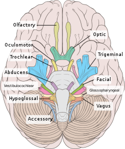
Inferior schematic view of the brain and brainstem showing the cranial nerves, numbered from olfactory to hypoglossal after the order in which they emerge.

Inferior view of the human brain showing the cranial nerves as visible on an autopsy specimen.
Traditionally, among humans there are considered to be twelve cranial nerves, numbered I-XII, all of which are paired. The cranial nerves arise directly from the central nervous system; the first two pairs, I and II, arise from the base of the forebrain, and the others, nerves III to XII, arise from the brainstem. Their naming scheme is given after their rostral-caudal orientation,[3][8] as, when viewing the brain and brainstem from below, they are often visible in their numeric order.
Unique anatomical terminology is used to describe the course of the cranial nerves. Like all nerves, the nerves have a nucleus, and a course within and outside of the brain. The course within the brain is known as the central course of the nerve, and the course after it has emerged from the brain as the peripheral course. The nerves are paired, which means that they occur on both the right and left sides. Some nerves cross from the right side to the left side, and this is known anatomically as decussating. If a nerve supplies a muscle, skin, or has another function on the same side of the body as where it originates, this is called an ipsilateral course. If the course is opposite to the nucleus of the nerve, this is known as a contralateral course.

Intercranial course

The cranial nerves serve to innervate the head and neck area,[8] including both somatic and autonomic motor innervation as well as sensory innervation. Together the cranial nerves supply sensory innervation of the special senses such as taste, vision; smell; hearing. They also supply afferens of the somatic senses: visceral sensation of the head and neck; balance, and proprioception combining vestibular perception with proprioceptive information from the head and neck.[9]
Distinct from the head the two cranial nerves: IX and X; the glossopharyngeal and vagus nerves innervate both motor and sensory synapses pertaining to abdominal organs (though not pelvic), as well as structures of the neck and chest.[3] Differentiating the cranial nerves from spinal nerves, the cranial nerves are not strictly bound to certain segments of the body (as in dermatomes), but rather organize after function, hence the innervated areas overlap significantly more than those of spinal nerves.[3] The accessory nerve XI, is considered either a cranial nerve or a spinal nerve which emanates level with the brain-stem.[3]
Similar to the dorsal root ganglia of the spinal nerves and parasympathetic ganglia of the sacral parasympathetic system, the sensory cranial nerves have a number of ganglia outside the central nervous system. The sensory ganglia are directly correspondent to dorsal root ganglia and are as known as cranial sensory ganglia, and found along the course of the cranial nerves, outside the brain[8] and skull. Sensory ganglia exist for nerves with sensory function; V, VII, VIII, IX, X.[3] There are also a number of parasympathetic cranial nerve ganglia, while sympathetic ganglia innervating the head and neck reside in the upper regions of the sympathetic trunk, and do not belong to the cranial nerves.[10]

Nuclei


The brainstem, with brainstem cranial nerve nuclei and tracts inside the brainstem shaded red to illustrate the deeper structures.
The nerve fibres in each nerve contain a nucleus either in the brainstem of the mesencephalon. The olfactory nerve (I) emerges from the olfactory bulb, and depending slightly on division the optic nerve (III) is deemed to emerge from the lateral geniculate nuclei. The rest of the nerves have their cell-bodies in the brainstem and thus originate in the brainstem.[10]
Olfactory nerve Olfactory bulb
Optic nerve Lateral geniculate nucleus
Oculomotor nerve Oculomotor nucleus
Edinger-Westphal nucleus
Trochlear nerve Trochlear nucleus
Trigeminal nerve Trigeminal nerve nuclei:
Mesencephalic nucleus
Principle sensory nucleus
Spinal trigeminal nucleus
Trigeminal motor nucleus
Abducens nerve Abducens nucleus
Facial nerve Facial motor nucleus
Superior salivatory nucleus
Vestibulocochlear nerve Vestibular nuclei
w. subnuclei
Cochlear nucleus
w. subnuclei
Glossopharyngeal nerve Solitary nucleus
Spinal nucleus of the trigeminal nerve
Lateral nucleus of vagal trigone.
Nucleus ambiguus
Inferior salivatory nucleus
Vagus nerve Dorsal nucleus of vagus nerve
Nucleus ambiguus
Solitary nucleus
Spinal trigeminal nucleus
Accessory nerve Spinal accessory nucleus
Nucleus ambiguus
Hypoglossal nerve Hypoglossal nucleus

Cranial nerve columns

Postero-lateral or dorso-lateral aspect of the brainstem showing sensory nuclei in blue.
Postero-lateral or dorso-lateral aspect of the brainstem showing motor nuclei in red.
Brainstem showing motor nuclei in red, and sensory nuclei in blue.
Brainstem nuclei with associated functions are often found in similar areas of the brainstem. These are also known as functional columns.[10] Functional columns are a result of the development of the spinal cord. Four columns of gray matter are present in the spinal cord during embryological development. Each column represents a different function, and contributes neurons to different nerves. Each nerve is innervated by neurons from one or more of the columns.[9]
As the spinal cord develops, there are four columns. These are the general somatic efferent column, the general visceral efferent column, the general visceral afferent column and general somatic afferent column. These columns also extend into the brainstem, but are divided into smaller pieces.[9] In the brainstem there are six columns.
Four 'general' columns contain fibres that supply sensation or control muscles:[11]
There are three additional columns which innervate organs and tissues developing from the branchial arches and inner ear. These are the following:

Extracranial course

With the exception of the olfactory nerve (I) and optic nerve (II), the cranial nerves emerge from the brainstem. The oculomotor nerve (III) and trochlear nerve (IV) emerge from the pons, the trigeminal (V), abducens (VI), facial (VII) and vestibulocochlea (VIII) from the midbrain, and the glossopharyngeal (IX), vagus (X), accessory (XI) and hypoglossal (XII) emerge from the medulla.[12]
The olfactory nerve (I) and optic nerve (II) emerge separately. The olfactory nerves emerge from the olfactory bulbs on either side of the crista galli, a bony projection below the frontal lobe, and the optic nerves (II) emerge from the lateral colliculus, swellings on either side of the temporal lobes of the brain.[12]

Exiting the skull

Exits of cranial nerves from the skull.[11]
Location Nerve
cribiform plate Olfactory nerve (I)
optic foramen Optic nerve (II)
superior orbital fissure Oculomotor (III)
Trochlear (IV)
Abducens (VI)
Trigeminal V1
(ophthalmic)
round foramen Trigeminal V2
(maxillary)
oval foramen Trigeminal V3
(mandibular)
internal auditory canal Facial (VII)
Vestibulocochlear (VIII)
jugular foramen Glossopharyngeal (IX)
Vagus (X)
Accessory (XI)
hypoglossal canal Hypoglossal (XII)
After emerging from the brainstem, the cranial nerves travel through the skull, but must leave this bony compartment in order to reach their destinations. Some nerves pass through unique holes in the skull, called foramina, as they travel to their destination. Other nerves pass through bony canals, longer canals enclosed by bone. These foramina and canals may contain more than one cranial nerve, and may also contain additional blood vessels.[11]
  • The olfactory nerve (I) passes through the cribiform plate, many small perforations in the ethmoid plate.
  • The optic nerve (II) passes through the optic foramen as it travels to the eye.
  • The oculomotor nerve (III), trochlear nerve (IV), abducens nerve (VI) and the opthalamic branch of the trigeminal nerve (V1) travel through the cavernous sinus into the superior orbital fissure, passing out of the skull into the orbit.
  • The maxillary division of the trigeminal nerve (V2) passes through the round foramen
  • The mandibular division of the trigeminal nerve (V3) passes through the oval foramen
  • The facial nerve (VII) and vestibulocochlear nerve (VIII) both pass through the internal auditory canal
The cranial nerves commonly enter and exit the skull together, for example nerves II, III, IV, V, and VI all pass through foramina near the pituitary fossa.[3]
The facial nerve enters the temporal bone at the internal acoustic meatus but exits the skull via the stylomastoid foramen while the vestibulocochlear nerve never actually exits the skull.

Ganglia

The cranial nerves give rise to a number of ganglia, collections of the cell bodies of neurons in the nerves that are outside of the brain. These ganglia are both parasympathetic and sensory ganglia.
The ganglion of the sensory nerves, which are similar in structure to the dorsal root ganglion of the spinal cord, include:[13]
Additional ganglia for nerves with parasympathetic function exist, and include the Ciliary ganglion of the oculomotor nerve (III), the pterygopalatine ganglion of the maxillary nerve (V2), the submandibular ganglion of the lingual nerve, a branch of the facial nerve (VII), and the otic ganglion of the glossopharyngeal nerve (IX).[14]

Course

The following images show the cranial nerves schematically showing their respective exits from the CNS or brain-stem (not including the optic nerve which does not leave the CNS), and their path, as well as conceptual innervation targets.

Summary

No. Name Sensory,
motor,
or both
Origin/Target Function
0 Terminal Purely sensory Lamina terminalis Animal research indicates that the terminal nerve is involved in the detection of pheromones.[15][unreliable medical source?][16]
I Olfactory Purely sensory Telencephalon Transmits the sense of smell from the nasal cavity.[17] Located in the olfactory foramina in the cribriform plate of the ethmoid bone.
II Optic Sensory Retinal ganglion cells Transmits visual signals from the retina of the eye to the brain.[18] Located in the optic canal.
III Oculomotor Mainly motor Anterior aspect of Midbrain Innervates the levator palpebrae superioris, superior rectus, medial rectus, inferior rectus, and inferior oblique, which collectively perform most eye movements. Also innervates the sphincter pupillae and the muscles of the ciliary body. Located in the superior orbital fissure.
IV Trochlear motor Dorsal aspect of Midbrain Innervates the superior oblique muscle, which depresses, rotates laterally, and intorts the eyeball. Located in the superior orbital fissure.
V Trigeminal Both sensory and motor Pons Receives sensation from the face and innervates the muscles of mastication.
Located in the;
superior orbital fissure (ophthalmic nerve - V1),
foramen rotundum (maxillary nerve - V2),
foramen ovale (mandibular nerve - V3).
VI Abducens Mainly motor Nuclei lying under the floor of the fourth ventricle
Pons
Innervates the lateral rectus, which abducts the eye. Located in the superior orbital fissure.
VII Facial Both sensory and motor Pons (cerebellopontine angle) above olive Provides motor innervation to the muscles of facial expression, posterior belly of the digastric muscle, stylohyoid muscle, and stapedius muscle. Also receives the special sense of taste from the anterior 2/3 of the tongue and provides secretomotorinnervation to the salivary glands (except parotid) and the lacrimal gland. Located in and runs through the internal acoustic canal to the facial canal and exits at the stylomastoid foramen.
VIII Vestibulocochlear
(also auditory,[19] acoustic,[19] or auditory-vestibular)
Mostly sensory Lateral to CN VII (cerebellopontine angle) Mediates sensation of sound, rotation, and gravity (essential for balance and movement). More specifically, the vestibular branch carries impulses for equilibrium and the cochlear branch carries impulses for hearing. Located in the internal acoustic canal.
IX Glossopharyngeal Both sensory and motor Medulla Receives taste from the posterior 1/3 of the tongue, provides secretomotor innervation to the parotid gland, and provides motor innervation to the stylopharyngeus. Some sensation is also relayed to the brain from the palatine tonsils. Located in the jugular foramen. This nerve is involved together with the vagus nerve in the gag reflex.
X Vagus Both sensory and motor Posterolateral sulcus of Medulla Supplies branchiomotorinnervation to most laryngeal and pharyngeal muscles (except the stylopharyngeus, which is innervated by the glossopharyngeal). Also provides parasympathetic fibers to nearly all thoracic and abdominal viscera down to the splenic flexure. Receives the special sense of taste from the epiglottis. A major function: controls muscles for voice and resonance and the soft palate. Symptoms of damage:dysphagia (swallowing problems), velopharyngeal insufficiency. Located in the jugular foramen. This nerve is involved (together with nerve IX) in the pharyngeal reflex or gag reflex.
XI Accessory
Sometimes:
cranial accessory
spinal accessory
Mainly motor Cranial and Spinal Roots Controls the sternocleidomastoid and trapezius muscles, and overlaps with functions of the vagus nerve (CN X). Symptoms of damage: inability to shrug, weak head movement. Located in the jugular foramen.
XII Hypoglossal Mainly motor Medulla Provides motor innervation to the muscles of the tongue (except for the palatoglossal muscle, which is innervated by the vagus nerve) and other glossal muscles. Important for swallowing (bolus formation) and speech articulation. Passes through the hypoglossal canal.

Function

Cranial nerve function is an important element in neurological examination, as specific dysfunction may indicate as to which portion of the brainstem is damaged. It is of clinical importance to know the path and origin of the cranial nerves, both intracranially as well as extracranially.[3]
The cranial nerves are often the first structures to be affected by different forms of brain injury such as hemorrhaging or tumors, partly because they are sensitive to compression.[10] Mononeuropathy of a cranial nerve may sometimes be the first symptom of an intracranial or skull base cancer.[20]

Smell (I)

Damage to the olfactory nerve can cause an inability to smell (anosmia), a distortion in the sense of smell (parosmia), or a distortion or lack of taste. Specific testing is performed when an individual perceives lack of taste or affected taste. The smell from each nostril is tested individually, and with consideration of airflow. Different substances are used, and these include coffee or soap. Using stronger smelling substances, for example ammonia, may lead to the activation of nociceptors of the trigeminal nerve.[21]

Vision (II)

Damage to the optic nerve affects vision. Vision is affected depending on the location of the lesion. A person may not be able to see things on their left or right side (homonymous hemianopsia), or may have difficulty seeing things on their outer visual fields (bitemporal hemianopsia) if the optic chiasm is involved.[1]:82 Vision may be tested using a number of different tests, examining the visual field, or by examining the cornea with a ophthalmoscope, using a process known as funduscopy. Visual field testing may be used to pin-point structural lesions in optic nerve, or further along the visual pathways.[21]

Eye movement (III, IV, VI)


Various deviations of the eyes due to abnormal function of the targets of the cranial nerves
Damage or lesion of nerves III, IV, or VI may affect the movement of the eye or pupil. Either both or one eye may be affected, and if both eyes are affected no double vision (diplopia) will occur. These nerves might be examined by observing how the eye follows an object in different directions. This object may be a finger or a pin, and may be moved at different directions to test for pursuit velocity.[21]
If the eyes do not work together, the most likely cause is damage to a specific cranial nerve or nuclei.[21]
    • Damage to the oculomotor nerve can cause double vision (diplopia) with lateral strabismus, and also ptosis and mydriasis.[1]:84 All but specific deviations may be due to damage in this nerve or any of the muscles it innervated, (though not internuclear ophthalmoplegia). Lesion may also lead to inability to open the eye, due to disrupted innervation of the levator palpebrae (unlike in Horner syndrome, which only results in a droopy eyelid. Individuals suffering from lesion or damage to the oculomotor nerve may compensate by tilting their heads to alleviate symptoms due to lack of control from oblique muscles when the eye is not adducted.[21]
    • Damage to the trochlear nerve can also cause diplopia with the eye adducted and elevated.[1]:84 The result will be an eye which can not move downwards or inwards properly (especially downwards when in an inward position). This is due to impairment in the superior oblique muscle innervated by the trochlear nerve.[21]
    • Damage to the abducens nerve can also result in diplopia with medial strabismus.[1]:84 This is due to impairment in the lateral rectus muscle innervated by the nerve.[21]

Facial sensation (V)

Conditions affecting the trigeminal nerve include trigeminal neuralgia,[10] cluster headache,[22] and trigeminal zoster.[10] Trigeminal neuraliga occurs later in life, from middle age onwards, most often after an age of 60; and is a condition associated with very strong pain distributed over the area innervated by the trigeminal nerve. Often the pain follows the distribution of the maxillary or mandibular nerve, (branches V2 and V3.[23] The trigeminal nerve is also present in the tendon reflexive jaw jerk. A reflex involving an induced twitch in muscles involved in closing the jaw when upon tapping on the jaw. A stronger reflex may be present if there is a supranuclear lesion of the trigeminal nerves motor nucleus, for example in pseudobulbar palsy.[23] In Parkinson's disease the trigeminal nerve is involved in the glabellar reflex which causes involuntary eye-blinking.

The facial nerve passes through the petrous temporal bone and internal auditory meatus after which it enters the facial canal to finally exit the stylomastoid foramen before passing through the parotid gland on its way to innervate the face.

Facial expression (VII)

Lesions of the facial nerve may manifest as facial palsy. This is where a person is unable to move the muscles on one or both sites of their face. If only the peripheral nerve itself is affected, this may cause Bell's palsy. Palsy that occurs is on the same side of the affected nerve. Central facial palsy will manifest in a similar fashion. If the nerve is damaged only on one side, a person will still be able to raise the eyebrows and crease the forehead on that side. That is because the frontalis muscle is innervated by both the left and the right cranial nerve. The effect is most often unilateral, and indicates contralateral damage or engagement of the cerebrum.[10]

Hearing and balance (VIII)

The vestibulocochlear nerve splits into the vestibular and cochlear nerve. The vestibular part is responsible for innervating the vestibules and semicircular canal of the inner ear, which transmits information about balance, and is an important component of the vestibuloocular reflex, which keeps the head stable and allows the eyes to track moving objects. The cochlear nerve transmits information from the cochlea, allowing sound to be heard.
When damaged:
  • The vestibular nerve may give rise to the sensation of spinning and dizziness, and may cause rotatory nystagmus. Function of the vestibular nerve may be tested through caloric stimulation.[21] Damage to the vestibulocochlear nerve can also present as repetitive and involuntary eye movements (nystagmus), particularly when looking in a horizontal plane.[21]
  • The cochlear nerve will cause partial or complete deafness in the affected ear.[21]

Oral sensation and taste (IX)


Deviating uvula due to cranial nerve X lesion.
The glossopharyngeal nerve is almost exclusively sensory in supplying five afferent nuclei of the brainstem, covering the oropharynx and back of the tongue with innervation.[24] Damage may result in difficulties swallowing.[21]

Vagus nerve (X)

Loss of function of the vagal nerve will lead to a loss of parasympathetic innervation to a very large number of structures. Of the major effects a rise in blood pressure and heart rate may occur. Isolated dysfunction of only the vagus nerve is rare, but can be diagnosed by a hoarse voice, due to dysfunction of the superior laryngeal nerve[10]
Testing of function may be performed by assessing ability to drink liquids. Choking on either saliva or liquids may indicated neurological damage to the vagal nerve.[21] Damage to the glossopharyngeal can be assessed by asking the subject to say "Ah" during phonation inspect to see if the uvula deviates. Positive sign indicative of unilateral damage occurs with finding of asymmetrically deviating uvula, towards the side with an intact or healthy nerve.[21]

Shoulder elevation and head-turning (XI)


Winged scapula may occur due to lesion of the spinal accessory.
Damage to the accessory nerve may lead to contralateral weakness in the trapezius, which can be tested by asking the subject to raise their shoulders or shrug, upon which the scapula will move out into a winged position if the nerve is damaged.[21] Weakness or an inability to elevate the scapula may be present, since the levator scapulae is alone in providing this function.[23]
There may also be weakness present of the sternocleidomastoid muscle, but as it received cortical innervation from the ipilateral side any damage will give rise to ipsilateral weakness.[21]

Tongue movement (XII)


A damaged hypoglossal nerve will result in an inability to stick the tongue out straight.
The hypoglossal nerve is unique in that it is innervated bilaterally from both hemispheres motor cortex. Damage to the nerve at lower motor neuron level may lead to fascinations of atrophy of the musculature of the tongue. The fasciculations of the tongue are sometimes said to look like a "bag of worms". Upper motor neuron damage will not lead to atrophy or fasciculations, but only weakness of the innervated muscles.[21]
When the nerve is damaged it will lead to unilateral weakness, and the tongue will upon being stuck out move towards the weaker or damaged side, as shown in the image.[21]

Clinical significance


Use of a Snellen chart to examine the optic nerve (II) may constitute one part of the cranial nerve examination.

Exam

Doctors, neurologists and other medical professionals may conduct a cranial nerve examination as part of a neurological examination to examine the cranial nerves. This is a highly formalised series of steps involving specific tests for each nerve.

Damage

Stroke

Stroke may damage the blood supply to areas of the nerves. It may also damage the areas of the brain that control the nerves. If there is a stroke of the midbrain, pons or medulla, various cranial nerves may be damaged, resulting in dysfunction and symptoms of a number of different syndromes.
Cavernous sinus thrombosis refers to a thrombus affecting the venous drainage from the cavernous sinus, which several cranial nerves pass through.

Compression

Nerves may be compressed because of increased intercranial pressure or tumour masses that presses against the nerves. For example, an optic glioma may impact on the optic nerve (II), and an acoustic neuroma may compress the facial nerve (VII) and vestibulocochlear nerve (VIII)

Inflammation

Inflammation can be a result of infection, such as viral causes, or can occur spontaneously. Inflammation is more common in some nerves than others. Spontaneous inflammation may result in a palsy of a nerve that self-resolves, such as Trochlea palsy. Inflammation of the facial nerve (VII) may result in Bell's palsy. Inflammation of the trigeminal nerve (V) may result in Trigeminal neuralgia, a phenominon in which the face is exquisitely tender.