Sunday 30 March 2014

Gross Structure and function of CSF

Cerebrospinal fluid (CSF) is a clear colorless bodily fluid found in the brain and spine. It is produced in the choroid plexus of the brain. It acts as a cushion or buffer for the brain's cortex, providing a basic mechanical and immunological protection to the brain inside the skull, and it serves a vital function in cerebral autoregulation of cerebral blood flow.
The CSF occupies the subarachnoid space (the space between the arachnoid mater and the pia mater) and the ventricular system around and inside the brain and spinal cord. It constitutes the content of the ventricles, cisterns, and sulci of the brain, as well as the central canal of the spinal cord.

Structure

CSF circulation

Production

The brain produces roughly 500 mL of cerebrospinal fluid per day. This fluid is constantly reabsorbed, so that only 100-160 mL is present at any one time. Ependymal cells of the choroid plexus produce more than two thirds of CSF. The choroid plexus is a venous plexus contained within the four ventricles of the brain, hollow structures inside the brain filled with CSF. The remainder of the CSF is produced by the surfaces of the ventricles and by the lining surrounding the subarachnoid space. [1] :764
Ependymal cells actively secrete sodium into the lateral ventricles. This creates osmotic pressure and draws water into the CSF space. Chloride, with a negative charge, maintains electroneutrality and moves with the positively-charged sodium. As a result, CSF contains a higher concentration of sodium and chloride than blood plasma, but less potassium, calcium and glucose and protein. [2]:519-520 [1]:764

Circulation

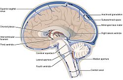
CSF circulates within the ventricular system of the brain. The majority of CSF is produced from within the lateral ventricles. From here, the CSF passes through the Interventricular foramina to the third ventricle, then the cerebral aqueduct to the fourth ventricle. The fourth ventricle is an outpouching on the posterior part of the brainstem. From the fourth ventricle, the fluid passes through the Foramen of Magendie (medial) and Foramen of Luschka (lateral) to enter the subarachnoid space, which covers the brain and spinal cord. [1]:764
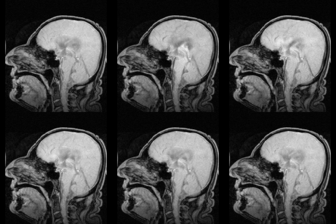
The CSF moves in a pulsatile manner throughout the CSF system with nearly zero net flow.[citation needed]

Reabsorption

MRI showing pulsation of CSF
It had been thought that CSF returns to the vascular system by entering the dural venous sinuses via the arachnoid granulations (or villi). However, some[3] have suggested that CSF flow along the cranial nerves and spinal nerve roots allow it into the lymphatic channels; this flow may play a substantial role in CSF reabsorbtion, in particular in the neonate, in which arachnoid granulations are sparsely distributed. The flow of CSF to the nasal submucosal lymphatic channels through the cribriform plate seems to be especially important.[4]
CSF is reabsorbed into venous sinus blood via arachnoid granulations.

Amount and constitution

Intracranial volumetric distribution of cerebrospinal fluid, blood, and brain parenchyma
Volumetric distribution of cerebrospinal fluid
The CSF contains approximately 0.3% plasma proteins, or approximately 15 to 40 mg/dL, depending on sampling site,[5] and it is produced at a rate of 500 ml/day. Since the subarachnoid space around the brain and spinal cord can contain only 135 to 150 ml, large amounts are drained primarily into the blood through arachnoid granulations in the superior sagittal sinus. Thus the CSF turns over about 3.7 times a day. This continuous flow into the venous system dilutes the concentration of larger, lipid-insoluble molecules penetrating the brain and CSF.[6]
CSF pressure, as measured by lumbar puncture (LP), is 10-18 cmH2O (8-15 mmHg or 1.1-2 kPa) with the patient lying on the side and 20-30cmH2O (16-24 mmHg or 2.1-3.2 kPa) with the patient sitting up.[7] In newborns, CSF pressure ranges from 8 to 10 cmH2O (4.4–7.3 mmHg or 0.78–0.98 kPa). Most variations are due to coughing or internal compression of jugular veins in the neck. When lying down, the cerebrospinal fluid as estimated by lumbar puncture is similar to the intracranial pressure.
There are quantitative differences in the distributions of a number of proteins in the CSF. In general, globular proteins and albumin are in lower concentration in ventricular CSF compared to lumbar or cisternal fluid.[8] The IgG index of cerebrospinal fluid is a measure of the immunoglobulin G content, and is elevated in multiple sclerosis. It is defined as IgG index = (IgGCSF / IgGserum ) / (albuminCSF / albuminserum).[9] A cutoff value has been suggested to be 0.73, with a higher value indicating presence of multiple sclerosis.[9]

Development

Around the third week of development, the embryo is a three-layered disc. The embryo is covered on the dorsal surface by a layer of cells called endoderm. In the middle of the dorsal surface of the embryo is a linear structure called the notochord. As the endoderm proliferates, the notochord is dragged into the middle of the developing embryo. The notochord becomes a canal within the embryo known as the neural canal. [10]
As the brain develops, by the fourth week of embryological development several swellings have formed within the embryo around the canal, near where the head will develop. These swellings represent different components of the central nervous system, and are three in number: the prosencephalon, mesencephalon and rhombencephalon.[10]
The developing forebrain surrounds the neural cord. As the forebrain develops, the neural cord within it becomes a ventricle, ultimately forming the lateral ventricles. Along the inner surface of both ventricles, the ventricular wall remains thin, and a choroid plexus develops, releasing CSF. The CSF quickly fills the neural canal.[10]

Function

CSF serves four primary purposes:
  1. Buoyancy: The actual mass of the human brain is about 1400 grams; however, the net weight of the brain suspended in the CSF is equivalent to a mass of 25 grams.[11] The brain therefore exists in neutral buoyancy, which allows the brain to maintain its density without being impaired by its own weight, which would cut off blood supply and kill neurons in the lower sections without CSF.[12]
  2. Protection: CSF protects the brain tissue from injury when jolted or hit. In certain situations such as auto accidents or sports injuries, the CSF cannot protect the brain from forced contact with the skull case, causing hemorrhaging, brain damage, and sometimes death.[12]
  3. Chemical stability: CSF flows throughout the inner ventricular system in the brain and is absorbed back into the bloodstream, rinsing the metabolic waste from the central nervous system through the blood–brain barrier. This allows for homeostatic regulation of the distribution of neuroendocrine factors, to which slight changes can cause problems or damage to the nervous system. For example, high glycine concentration disrupts temperature and blood pressure control, and high CSF pH causes dizziness and syncope.[12] To use Davson's term, the CSF has a "sink action" by which the various substances formed in the nervous tissue during its metabolic activity diffuse rapidly into the CSF and are thus removed into the bloodstream as CSF is absorbed.[13]
  4. Prevention of brain ischemia: The prevention of brain ischemia is made by decreasing the amount of CSF in the limited space inside the skull. This decreases total intracranial pressure and facilitates blood perfusion.

Friday 28 March 2014

Brain Lobes & their functions.

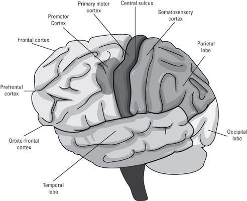
The neocortex is divided into four major lobes: the frontal lobe, the parietal lobe, the temporal lobe, and occipital lobe. These lobes are further divided into different regions. The frontal lobes are involved with control of movement, from stimulation of individual muscles to abstract planning about what to do.
The parietal lobe processes visual, auditory and touch information. The temporal lobe is the primary area for early auditory processing and a high level visual processing area. The occipital lobe processes visual information and sends it to the parietal and temporal lobes.
The four lobes and the regions within each.

The frontal lobe of the brain

The frontal lobe is concerned with executing behavior. This ranges from the control of individual muscles in the primary motor cortex to high level abstract planning about what to do. The frontal lobes are divided into different areas:
  • The prefrontal cortex: In humans, the prefrontal cortex takes up the majority of the frontal lobe. The prefrontal cortex is crucial for the performance of almost all skills requiring intelligence. The prefrontal cortex tends to be larger in primates than other mammals, and it’s larger in humans than in other primates. This is correlated with the amount of high level planning done by members of different species.
    Most mammals operate mostly on instinct and don’t live in complexly differentiated social groups. Primates, on the other hand, have complex male and female hierarchies and may hatch plots against each other that span years of planning. Humans build tools, modify their environments for their own purposes, and have specific relationships with up to hundreds of other individuals (and this was even before Facebook).
  • The orbitofrontal cortex: This area is the anterior and medial part of the prefrontal cortex. The orbitofrontal cortex is essential for risk and reward assessment and for what might be called moral judgment. Patients with damage to this area may have normal or superior intelligence as assessed by IQ tests but lack even a rudimentary concept of manners or appropriate actions in social contexts; they also lose almost all risk aversion despite clear knowledge of bad consequences.
  • Primary motor cortex: The primary motor cortex is the strip of brain area just anterior to the central sulcus, the most posterior portion of the frontal lobe. The brain can take direct control of the muscles from the spinal cord. It does this through projections from the primary motor cortex. Neurons in the primary motor cortex travel down the spinal cord and synapse on the same motor neurons that mediate reflexes. In theory, this direct control allows far more flexibility and adaptability.
  • Premotor cortex: The job of the premotor cortex is to consciously monitor movement sequences, using sensory feedback. After the basal ganglia and prefrontal cortex select the goal, the premotor cortex coordinates the steps to reach that goal. Activity in the premotor cortex helps you learn what to pay attention to while you perform a complicated motor sequence and what to do when you get stuck at some particular point.
Think of the frontal cortex as “polarized” from anterior (front) to posterior (back). Farthest back, at the central sulcus, are neural wires going almost directly to muscles. In front of that are areas that organize and sequence movements. In front of that are abstract planning levels. At these abstract levels, for example, you select from a variety of different strategies that may involve completely different muscles, muscles sequences, or, as in the tennis shot, the decision to not move at all.

The brain's parietal lobe

The parietal lobe contains neurons that receive sensory information from the skin and tongue, and processes sensory information from the ears and eyes that are received in other lobes. The major sensory inputs from the skin (touch, temperature, and pain receptors) relay through the thalamus to parietal lobe.

The occipital lobe

The occipital lobe processes visual input that is sent to the brain from the retinas. The retinas project onto the posterior pole of the occipital lobe, called V1 (for visual area one), so that activity in different areas of V1 is related to whatever is in the image around your current point of gaze.
The four lobes and the regions within each.Subareas beyond V1 specialize in visual tasks such as color detection, depth perception, and motion detection. The sense of vision is further processed by projections from these higher occipital lobe areas to other areas in the parietal and temporal lobes, but this processing is dependent on early processing by the occipital lobe. (Researchers know this because damage to V1 causes blindness in that part of the visual field that projects there.)
The fact that the visual system gets an entire lobe for processing emphasizes the importance of high visual acuity and processing among our senses.

The temporal lobe

The brain's temporal lobe combines auditory and visual information. The superior (upper) and medial (central) aspect of the temporal lobe receives auditory input from the part of the thalamus that relays information from the ears. The inferior (lower) part of the temporal lobe does visual processing for object and pattern recognition. The medial and anterior parts of the temporal lobe are involved in very high-order visual recognition (being able to recognize faces, for example), as well as recognition depending on memory.

Wednesday 26 March 2014

Neurons

A neuron  is an electrically  excitable cell  that processes and transmits information through electrical and chemical signals. These signals between neurons occur via synapses, specialized connections with other cells. Neurons can connect to each other to form neural networks. Neurons are the core components of the nervous system, which includes the brain, and spinal cord of the central nervous system (CNS), and the ganglia of the peripheral nervous system (PNS). Specialized types of neurons include: sensory neurons which respond to touch, sound, light and all other stimuli affecting the cells of the sensory organs, that then send signals to the spinal cord and brain; motor neurons that receive signals from the brain and spinal cord, to cause muscle contractions, and affect glandular outputs, and interneurons which connect neurons to other neurons within the same region of the brain or spinal cord, in neural networks.


A typical neuron possesses a cell body (soma), dendrites, and an axon. The term neurite is used to describe either a dendrite or an axon, particularly in its undifferentiated stage. Dendrites are thin structures that arise from the cell body, often extending for hundreds of micrometres and branching multiple times, giving rise to a complex "dendritic tree". An axon is a special cellular extension that arises from the cell body at a site called the axon hillock and travels for a distance, as far as 1 meter in humans or even more in other species. The cell body of a neuron frequently gives rise to multiple dendrites, but never to more than one axon, although the axon may branch hundreds of times before it terminates. At the majority of synapses, signals are sent from the axon of one neuron to a dendrite of another. There are, however, many exceptions to these rules: neurons that lack dendrites, neurons that have no axon, synapses that connect an axon to another axon or a dendrite to another dendrite, etc.


All neurons are electrically excitable, maintaining voltage gradients across their membranes by means of metabolically driven ion pumps, which combine with ion channels embedded in the membrane to generate intracellular-versus-extracellular concentration differences of ions such as sodium, potassium, chloride, and calcium. Changes in the cross-membrane voltage can alter the function of voltage-dependent ion channels. If the voltage changes by a large enough amount, an all-or-none electrochemical pulse called an action potential is generated, which travels rapidly along the cell's axon, and activates synaptic connections with other cells when it arrives.
Neurons do not undergo cell division. In most cases, neurons are generated by special types of stem cells. A type of glial cell, called astrocytes (named for being somewhat star-shaped), have also been observed to turn into neurons by virtue of the stem cell characteristic pluripotency. In humans, neurogenesis largely ceases during adulthood—but in two brain areas, the hippocampus and olfactory bulb, there is strong evidence for generation of substantial numbers of new neurons

Monday 24 March 2014

Basic structure and function of Brain

The nervous system is your body's decision and communication center. The central nervous system (CNS) is made of the brain and the spinal cord and the peripheral nervous system (PNS) is made of nerves. Together they control every part of your daily life, from breathing and blinking to helping you memorize facts for a test. Nerves reach from your brain to your face, ears, eyes, nose, and spinal cord... and from the spinal cord to the rest of your body. Sensory nerves gather information from the environment, send that info to the spinal cord, which then speed the message to the brain. The brain then makes sense of that message and fires off a response. Motor neurons deliver the instructions from the brain to the rest of your body. The spinal cord, made of a bundle of nerves running up and down the spine, is similar to a superhighway, speeding messages to and from the brain at every second.


The brain is made of three main parts: the forebrain, midbrain, and hindbrain. The forebrain consists of the cerebrum, thalamus, and hypothalamus (part of the limbic system). The midbrain consists of the tectum and tegmentum. The hindbrain is made of the cerebellum, pons and medulla. Often the midbrain, pons, and medulla are referred to together as the brainstem.

The Cerebrum: The cerebrum or cortex is the largest part of the human brain, associated with higher brain function such as thought and action. The cerebral cortex is divided into four sections, called "lobes": the frontal lobe, parietal lobe, occipital lobe, and temporal lobe. Here is a visual representation of the cortex:
What do each of these lobes do?
  • Frontal Lobe- associated with reasoning, planning, parts of speech, movement, emotions, and problem solving
  • Parietal Lobe- associated with movement, orientation, recognition, perception of stimuli
  • Occipital Lobe- associated with visual processing
  • Temporal Lobe- associated with perception and recognition of auditory stimuli, memory, and speech
Note that the cerebral cortex is highly wrinkled. Essentially this makes the brain more efficient, because it can increase the surface area of the brain and the amount of neurons within it. We will discuss the relevance of the degree of cortical folding (or gyrencephalization) later.


A deep furrow divides the cerebrum into two halves, known as the left and right hemispheres. The two hemispheres look mostly symmetrical yet it has been shown that each side functions slightly different than the other. Sometimes the right hemisphere is associated with creativity and the left hemispheres is associated with logic abilities. The corpus callosum is a bundle of axons which connects these two hemispheres.
Nerve cells make up the gray surface of the cerebrum which is a little thicker than your thumb. White nerve fibers underneath carry signals between the nerve cells and other parts of the brain and body.
The neocortex occupies the bulk of the cerebrum. This is a six-layered structure of the cerebral cortex which is only found in mammals. It is thought that the neocortex is a recently evolved structure, and is associated with "higher" information processing by more fully evolved animals (such as humans, primates, dolphins, etc).


The Cerebellum: The cerebellum, or "little brain", is similar to the cerebrum in that it has two hemispheres and has a highly folded surface or cortex. This structure is associated with regulation and coordination of movement, posture, and balance.
The cerebellum is assumed to be much older than the cerebrum, evolutionarily. What do I mean by this? In other words, animals which scientists assume to have evolved prior to humans, for example reptiles, do have developed cerebellums. However, reptiles do not have neocortex


Limbic System: The limbic system, often referred to as the "emotional brain", is found buried within the cerebrum. Like the cerebellum, evolutionarily the structure is rather old.
This system contains the thalamus, hypothalamus, amygdala, and hippocampus. Here is a visual representation of this system, from a midsagittal view of the human brain:


  :


Brain Stem: Underneath the limbic system is the brain stem. This structure is responsible for basic vital life functions such as breathing, heartbeat, and blood pressure. Scientists say that this is the "simplest" part of human brains because animals' entire brains, such as reptiles


Sunday 23 March 2014


ENTIRE CENTRAL NERVOUS SYSTEM - A REVIEW

Together, the brain and spinal cord form the central nervous system. This complex system is part of everything we do. It controls the things we choose to do -- like walk and talk -- and the things our body does automatically -- like breathe and digest food. The central nervous system is also involved with our senses -- seeing, hearing, touching, tasting, and smelling -- as well as our emotions, thoughts, and memory.
The brain is a soft, spongy mass of nerve cells and supportive tissue. It has three major parts: the cerebrum, the cerebellum, and the brain stem. The parts work together, but each has special functions.


The cerebrum, the largest part of the brain, fills most of the upper skull. It has two halves called the left and right cerebral hemispheres. The cerebrum uses information from our senses to tell us what's going on around us and tells our body how to respond. The right hemisphere controls the muscles on the left side of the body, and the left hemisphere controls the muscles on the right side of the body. This part of the brain also controls speech and emotions as well as reading, thinking, and learning.
The cerebellum, under the cerebrum at the back of the brain, controls balance and complex actions like walking and talking.
The brain stem connects the brain with the spinal cord. It controls hunger and thirst and some of the most basic body functions, such as body temperature, blood pressure, and breathing.


The brain is protected by the bones of the skull and by a covering of three thin membranes called meninges. The brain is also cushioned and protected by cerebrospinal fluid. This watery fluid is produced by special cells in the four hollow spaces in the brain, called ventricles. It flows through the ventricles and in spaces between the meninges. Cerebrospinal fluid also brings nutrients from the blood to the brain and removes waste products from the brain.
The spinal cord is made up of bundles of nerve fibers. It runs down from the brain through a canal in the center of the bones of the spine. These bones protect the spinal cord. Like the brain, the spinal cord is covered by the meninges and cushioned by cerebrospinal fluid.
Spinal nerves connect the brain with the nerves in most parts of the body. Other nerves go directly from the brain to the eyes, ears, and other parts of the head. This network of nerves carries messages back and forth between the brain and the rest of the body

Saturday 22 March 2014

Human Brain a miracle of god


The  human brain is most complicated creation of God. even scientists , medical research sometimes puzzle to understand and cure it. Science and human being, with all their resources, cant fully understand due its complication, mystery of formation.


How a small fish can measure the depth of ocean. whatever we were able to find about mystery of  bhagwanji is very tiny.it is impossible to find all mystery. t is human brain that created science...; science is a puppet in the hands of human beings.....; no wonder of science can ever reveal the mystery of God's best creation...''human brain''..........