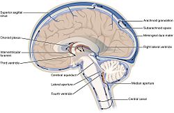
Cerebrospinal fluid (CSF) is a clear colorless bodily fluid found in the brain and spine. It is produced in the choroid plexus of the brain. It acts as a cushion or buffer for the brain's cortex, providing a basic mechanical and immunological protection to the brain inside the skull, and it serves a vital function in cerebral autoregulation of cerebral blood flow.
The CSF occupies the subarachnoid space (the space between the arachnoid mater and the pia mater) and the ventricular system around and inside the brain and spinal cord. It constitutes the content of the ventricles, cisterns, and sulci of the brain, as well as the central canal of the spinal cord.
Ependymal cells actively secrete sodium into the lateral ventricles. This creates osmotic pressure and draws water into the CSF space. Chloride, with a negative charge, maintains electroneutrality and moves with the positively-charged sodium. As a result, CSF contains a higher concentration of sodium and chloride than blood plasma, but less potassium, calcium and glucose and protein. [2]:519-520 [1]:764
The CSF moves in a pulsatile manner throughout the CSF system with nearly zero net flow.[citation needed]
CSF is reabsorbed into venous sinus blood via arachnoid granulations.
CSF pressure, as measured by lumbar puncture (LP), is 10-18 cmH2O (8-15 mmHg or 1.1-2 kPa) with the patient lying on the side and 20-30cmH2O (16-24 mmHg or 2.1-3.2 kPa) with the patient sitting up.[7] In newborns, CSF pressure ranges from 8 to 10 cmH2O (4.4–7.3 mmHg or 0.78–0.98 kPa). Most variations are due to coughing or internal compression of jugular veins in the neck. When lying down, the cerebrospinal fluid as estimated by lumbar puncture is similar to the intracranial pressure.
There are quantitative differences in the distributions of a number of proteins in the CSF. In general, globular proteins and albumin are in lower concentration in ventricular CSF compared to lumbar or cisternal fluid.[8] The IgG index of cerebrospinal fluid is a measure of the immunoglobulin G content, and is elevated in multiple sclerosis. It is defined as IgG index = (IgGCSF / IgGserum ) / (albuminCSF / albuminserum).[9] A cutoff value has been suggested to be 0.73, with a higher value indicating presence of multiple sclerosis.[9]
As the brain develops, by the fourth week of embryological development several swellings have formed within the embryo around the canal, near where the head will develop. These swellings represent different components of the central nervous system, and are three in number: the prosencephalon, mesencephalon and rhombencephalon.[10]
The developing forebrain surrounds the neural cord. As the forebrain develops, the neural cord within it becomes a ventricle, ultimately forming the lateral ventricles. Along the inner surface of both ventricles, the ventricular wall remains thin, and a choroid plexus develops, releasing CSF. The CSF quickly fills the neural canal.[10]
The CSF occupies the subarachnoid space (the space between the arachnoid mater and the pia mater) and the ventricular system around and inside the brain and spinal cord. It constitutes the content of the ventricles, cisterns, and sulci of the brain, as well as the central canal of the spinal cord.
Structure
Production
The brain produces roughly 500 mL of cerebrospinal fluid per day. This fluid is constantly reabsorbed, so that only 100-160 mL is present at any one time. Ependymal cells of the choroid plexus produce more than two thirds of CSF. The choroid plexus is a venous plexus contained within the four ventricles of the brain, hollow structures inside the brain filled with CSF. The remainder of the CSF is produced by the surfaces of the ventricles and by the lining surrounding the subarachnoid space. [1] :764Ependymal cells actively secrete sodium into the lateral ventricles. This creates osmotic pressure and draws water into the CSF space. Chloride, with a negative charge, maintains electroneutrality and moves with the positively-charged sodium. As a result, CSF contains a higher concentration of sodium and chloride than blood plasma, but less potassium, calcium and glucose and protein. [2]:519-520 [1]:764
Circulation
See also: Ventricular system
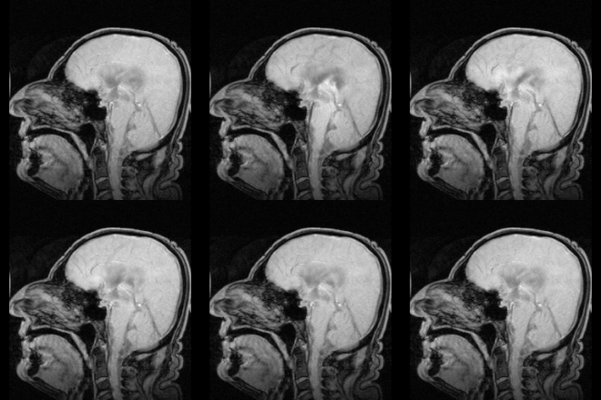
CSF circulates within the ventricular system of the brain. The majority of CSF is produced from within the lateral ventricles. From here, the CSF passes through the Interventricular foramina to the third ventricle, then the cerebral aqueduct to the fourth ventricle. The fourth ventricle is an outpouching on the posterior part of the brainstem. From the fourth ventricle, the fluid passes through the Foramen of Magendie (medial) and Foramen of Luschka (lateral) to enter the subarachnoid space, which covers the brain and spinal cord. [1]:764The CSF moves in a pulsatile manner throughout the CSF system with nearly zero net flow.[citation needed]
Reabsorption
It had been thought that CSF returns to the vascular system by entering the dural venous sinuses via the arachnoid granulations (or villi). However, some[3] have suggested that CSF flow along the cranial nerves and spinal nerve roots allow it into the lymphatic channels; this flow may play a substantial role in CSF reabsorbtion, in particular in the neonate, in which arachnoid granulations are sparsely distributed. The flow of CSF to the nasal submucosal lymphatic channels through the cribriform plate seems to be especially important.[4]CSF is reabsorbed into venous sinus blood via arachnoid granulations.
Amount and constitution
The CSF contains approximately 0.3% plasma proteins, or approximately 15 to 40 mg/dL, depending on sampling site,[5] and it is produced at a rate of 500 ml/day. Since the subarachnoid space around the brain and spinal cord can contain only 135 to 150 ml, large amounts are drained primarily into the blood through arachnoid granulations in the superior sagittal sinus. Thus the CSF turns over about 3.7 times a day. This continuous flow into the venous system dilutes the concentration of larger, lipid-insoluble molecules penetrating the brain and CSF.[6]CSF pressure, as measured by lumbar puncture (LP), is 10-18 cmH2O (8-15 mmHg or 1.1-2 kPa) with the patient lying on the side and 20-30cmH2O (16-24 mmHg or 2.1-3.2 kPa) with the patient sitting up.[7] In newborns, CSF pressure ranges from 8 to 10 cmH2O (4.4–7.3 mmHg or 0.78–0.98 kPa). Most variations are due to coughing or internal compression of jugular veins in the neck. When lying down, the cerebrospinal fluid as estimated by lumbar puncture is similar to the intracranial pressure.
There are quantitative differences in the distributions of a number of proteins in the CSF. In general, globular proteins and albumin are in lower concentration in ventricular CSF compared to lumbar or cisternal fluid.[8] The IgG index of cerebrospinal fluid is a measure of the immunoglobulin G content, and is elevated in multiple sclerosis. It is defined as IgG index = (IgGCSF / IgGserum ) / (albuminCSF / albuminserum).[9] A cutoff value has been suggested to be 0.73, with a higher value indicating presence of multiple sclerosis.[9]
Development
Around the third week of development, the embryo is a three-layered disc. The embryo is covered on the dorsal surface by a layer of cells called endoderm. In the middle of the dorsal surface of the embryo is a linear structure called the notochord. As the endoderm proliferates, the notochord is dragged into the middle of the developing embryo. The notochord becomes a canal within the embryo known as the neural canal. [10]As the brain develops, by the fourth week of embryological development several swellings have formed within the embryo around the canal, near where the head will develop. These swellings represent different components of the central nervous system, and are three in number: the prosencephalon, mesencephalon and rhombencephalon.[10]
The developing forebrain surrounds the neural cord. As the forebrain develops, the neural cord within it becomes a ventricle, ultimately forming the lateral ventricles. Along the inner surface of both ventricles, the ventricular wall remains thin, and a choroid plexus develops, releasing CSF. The CSF quickly fills the neural canal.[10]
Function
CSF serves four primary purposes:- Buoyancy: The actual mass of the human brain is about 1400 grams; however, the net weight of the brain suspended in the CSF is equivalent to a mass of 25 grams.[11] The brain therefore exists in neutral buoyancy, which allows the brain to maintain its density without being impaired by its own weight, which would cut off blood supply and kill neurons in the lower sections without CSF.[12]
- Protection: CSF protects the brain tissue from injury when jolted or hit. In certain situations such as auto accidents or sports injuries, the CSF cannot protect the brain from forced contact with the skull case, causing hemorrhaging, brain damage, and sometimes death.[12]
- Chemical stability: CSF flows throughout the inner ventricular system in the brain and is absorbed back into the bloodstream, rinsing the metabolic waste from the central nervous system through the blood–brain barrier. This allows for homeostatic regulation of the distribution of neuroendocrine factors, to which slight changes can cause problems or damage to the nervous system. For example, high glycine concentration disrupts temperature and blood pressure control, and high CSF pH causes dizziness and syncope.[12] To use Davson's term, the CSF has a "sink action" by which the various substances formed in the nervous tissue during its metabolic activity diffuse rapidly into the CSF and are thus removed into the bloodstream as CSF is absorbed.[13]
- Prevention of brain ischemia: The prevention of brain ischemia is made by decreasing the amount of CSF in the limited space inside the skull. This decreases total intracranial pressure and facilitates blood perfusion.


No comments:
Post a Comment